A corporate working women who has a daughter of 15 years was diagnosed with Cancer of Right Breast in the year 2018. She underwent Lumpectomy/radiotherapy, and had received 19 cycles of chemotherapy (last on 30/12/2019)
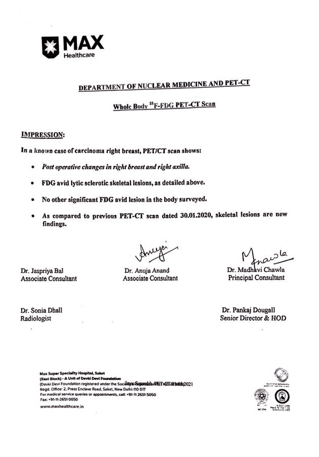
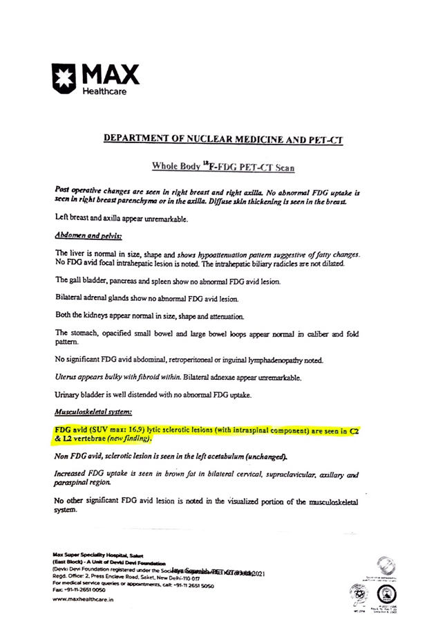
She approached us in mid-January 2021 with relapse and metastasis. Her PET-CT scandone on 28/12/2020 showed metastasis of tumour. Lytic sclerotic lesionsare seen in C2 and L2 vertebrae of size 16.9.(pic 1,2)
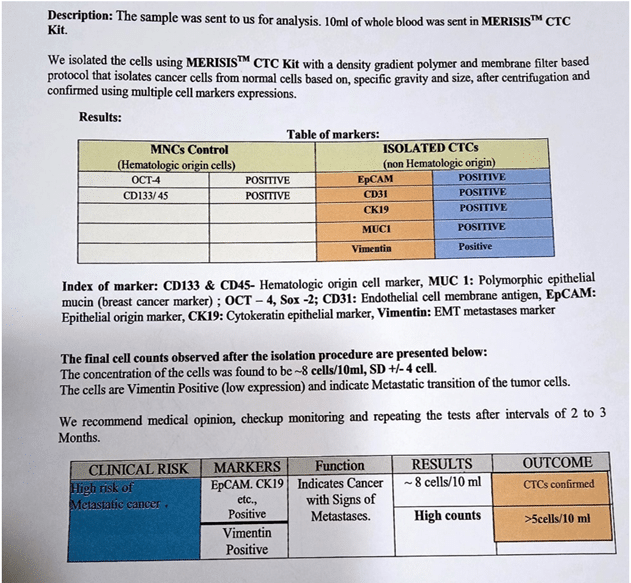
We did investigations to check her circulating tumour cell load with the help of special investigation and it stated high risk of metastatic cancer with high counts of cancer cell i.e. ~8 cells\10 ml.
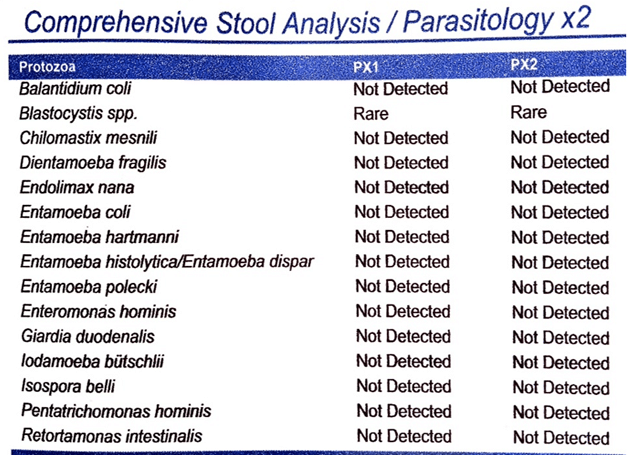
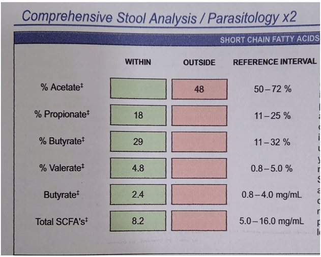
Other investigations like Stool comprehensive tests showed imbalanced gut flora and parasitic infestations (pic 4). Lowelastase which is an indicator of pancreatic exocrine insufficiency was found.Also Secretary IgA was abnormally high.
Stool Comprehensive report
Stool Comprehensive report
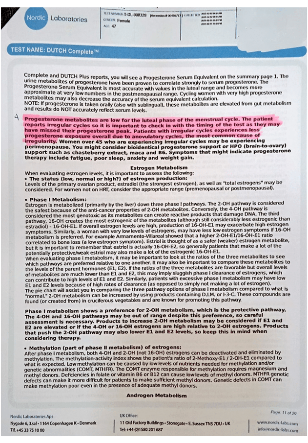
Her DUTCH testthat is a specialized hormone test revealed very low levels of progesterone (pic 6). Blood investigations showed her to be highly positive for EBV.
After investigations she was put on anti-cancer diet protocol, nutraceutical to help cope with identified nutritional deficiencies, intravenous high dose vitamin C therapy,ozonized saline drips and bio identical progesterone was prescribed. Intensive gut healing, liver detox to enhance hormone detoxification was implemented and following were the results:
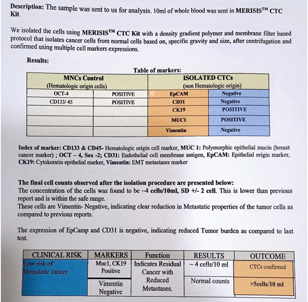
Her CTC report showed low risk of metastatic cancer (20/05/2021) and her cancer cells dropped down to ~4 cells/10ml.
CTC Report after treatment
After 6 months of continuous treatment following was the outcome:
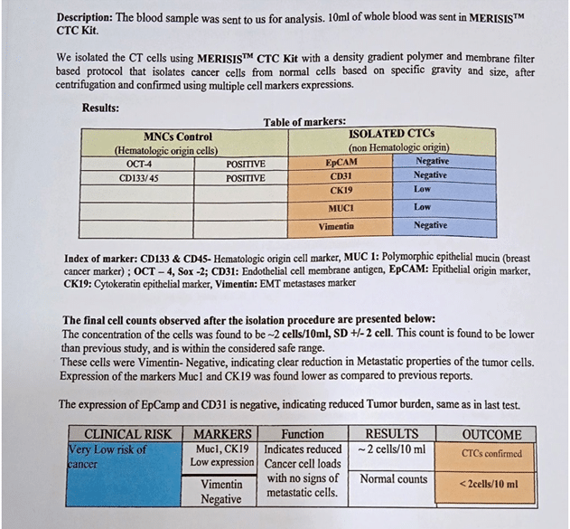
Massive fall in count of cancer cells was found ~2cells/10 ml and indicated reduced cancer cell load with no significant FDG of metastatic cells.
CTC Report after 6 months of continuous treatment
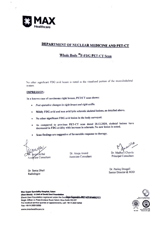
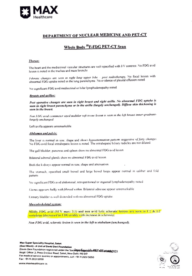
PET CT scan (03/05/2021) showed no new lesions, and lytic sclerotic size dropped to 3.1.
PET CT scan after treatment
PET CT scan after treatment
The reports showed favourable response to the treatment.